200以上 pancreas histology exocrine cells 149762
Exocrine Pancreas Histology Pancreatic acini are located within lobules At the base of each of the pancreatic acinar cells are abundant rough endoplasmic reticulum (RER) to produce proteins for The morphology of the exocrine secretory unit of the pancreas, ie the pancreatic acinus, is reviewed The histological features of the acini and their relation with the duct system are described The acinar three‐dimensional architecture was studied by means of different ultrastructural techniques, some of which are complementaryThe pancreas is both an exocrine (compound acinar) and an endocrine (islets of Langerhans) gland The exocrine pancreas releases alkaline secretions, containing enzymes, into the duodenum that aid in food digestion and neutralize the acidity of chyme leaving the stomach
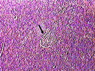
Pancreas
Pancreas histology exocrine cells
Pancreas histology exocrine cells-Including the uncinate portion;Pancreatic Histology Endocrine Tissue If you examine a section of pancreas at low magnification, you will undoubtedly observe distinctive areas of pale staining embedded within the exocrine tissue of lobules (marked with a red * below) These are the Islets of Langerhans, the endocrine component of the pancreas


Pancreas Histology Boyar
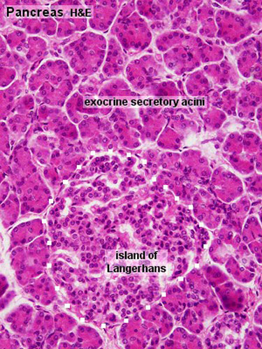
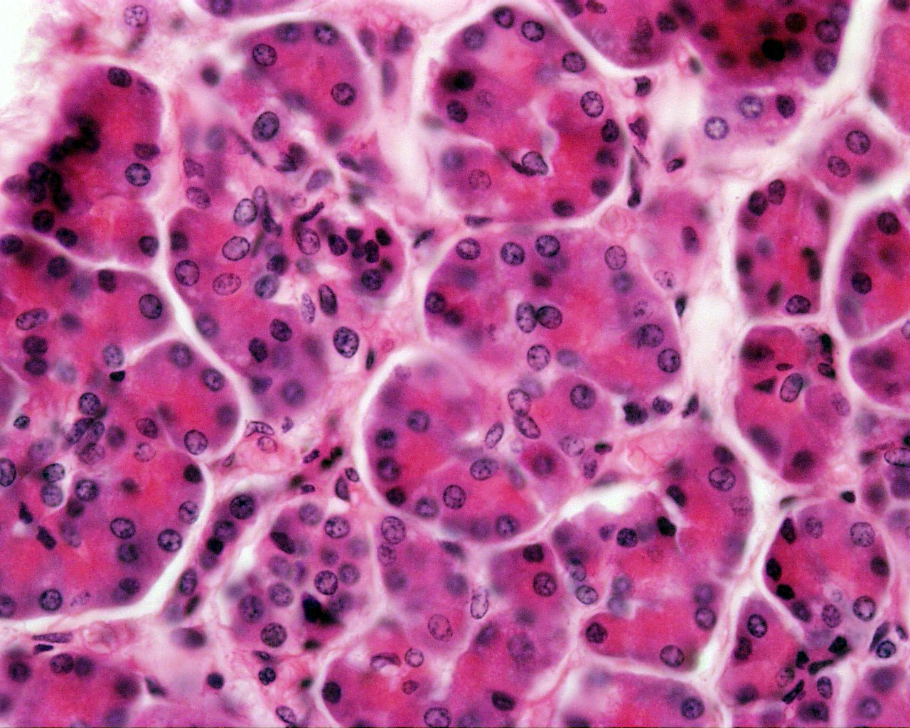
Pancreas Acini of the exocrine pancreas demonstrate the polarity of exocrine secretory cells Rough endoplasmic reticulum is evident at the periphery of each acinar cell;As in salivary glands, intercalated ductal cells in the pancreas contribute bicarbonate ions (sodium and water follow passively) to the exocrine secretory product However, unlike salivary glands, there are no striated ducts in the pancreas to recover sodium, so the final product is rich in both sodium and bicarbonate (as opposed to saliva in which the sodium content is about one tenthThe endocrine pancreas that secretes insulin and glucogon is more lightly stained and its cells cluster to form the Islets of Langerhans The rest of the image consists of the exocrine pancreas that produces several enzymes critical for digestion and adsorption of food Note how the cells of the exocrine pancreas form small clusters or acini
Histology of the Exocrine Pancreas Acini 1422 Ultrastructure of the Exocrine Pancreas 1423 Development of the Pancreas Parenchymal cells proliferating from this diverticulum pass into mesenchyme of the septum transversum and occupy meshes of a network of capillaries related to vitelline and umbilical vessels The exocrine secretory part consists of serous acini (known as pancreatic acini), whereas the islets of Langerhans form the endocrine part The major part of pancreas corresponds to the exocrine part (99 % in humans) The serous exocrine cells show a rounded nucleus and dark cytoplasm In pancreas histology of animal, you will find the both endocrine and exocrine parts The exocrine part forms the major portion of pancreas and consists of closely packed serous acini with zymogenic cells Again, the endocrine part consists of pancreatic islets of Langerhans which is located within the masses of serous acini
The pancreas is divided into lobules by connective tissue septae Lobules are composed largely of grapelike clusters of exocrine cells called acini, which sExocrine pancreatic status is directly linked to genotype (Kerem et al 1990;Histology in the Pancreas and MRI Imaging Biomarkers Temel Tirkes, Indiana University Organoid and Pancreas on Chip Alex Kleger, MD, Ulm University 3D Imaging Study and Mapped Changes in Innervation in Type 2 Diabetes Sarah Stanley, MBBCh, PhD, Mount Sinai Pancreas Slices to Study Interactions between Immune Cells and Islets
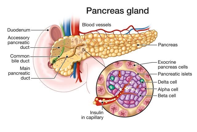


Histology Of Pancreas Structure Of Islets Of Langerhans Insulin Function Metabolism Science Online


Liver Gall Bladder Pancreas
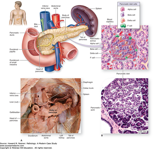
Pancreas Has both exocrine and endocrine functions Most of the pancreas comprises exocrine cells, called acini, that secrete digestive enzymes;Kristidis et al 1992) Analysis of particular CFTR mutations in patients with these pancreatic phenotypes (PI vs PS) revealed two categories of alleles "severe" and "mild" Patients homozygous or compound heterozygous for severe alleles belonging to classes I, II, III, or VI confer pancreatic insufficiency, Histology serous ductal / acinar (exocrine cells) gland with tubuloalveolar arrangement with interspersed islets of Langerhans (endocrine cells) Terminology Anatomically, from right to left, the pancreas is divided into the caput (head), collum (neck), corpus (body) and cauda (tail) Pancreatic secretion is drained by the main pancreatic duct


Digestive The Histology Guide


1
The pancreas is a twoheaded organ, not only in origin but also in function In origin, the pancreas develops from two separate primordia In function, the organ has both endocrine function in relation to regulating blood glucose (and also other hormone secretions) and gastrointestinal function as an exocrine (digestive) organ, see exocrine pancreas Exocrine vs Endocrine Fundamentally, the difference between exocrine and endocrine is exocrine secretes (via ducts) to an external surface or body cavity endocrine secretes within an enclosed system eg the bloodstream As the acini of the pancreas secrete into a network of ducts, this gives this organ an exocrine functionDescribe the endocrine function of the pancreas involves the release of hormones that regulate carbohydrate metabolism;


Osmosis The Pancreas Is A Large Mostly Retroperitoneal Organ That Has Important Exocrine And Endocrine Functions The Exocrine Digestive Enzymes Secreted By The Pancreas Include Lipase Amylase Pancreatin Trypsin And Chymotrypsin
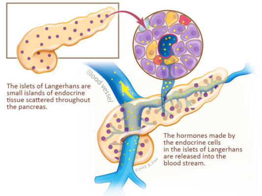


Pancreas Function Pancreatic Cancer Johns Hopkins Pathology
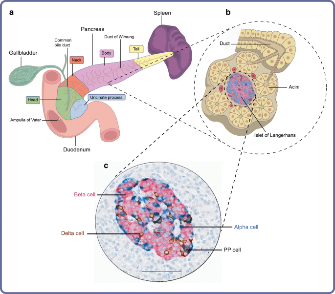
The pancreas contains two sets of cells exocrine cells that secrete enzymes via ducts into the intestine, and endocrine cells that secrete hormones into the bloodstream The exocrine cells are small and arranged in spherical secretory units called acini On a slide, one acinus would look something like a pie cut into several pieces4 rows The pancreas is both an exocrine accessory digestive organ and a hormone secreting endocrineIn humans, it is located in the abdomen behind the stomach and functions as a gland The pancreas has both an endocrine and a digestive exocrine function As an endocrine gland, it functions mostly to regulate blood sugar levels, secreting the hormones insulin, glucagon, somatostatin, and pancreatic polypeptide
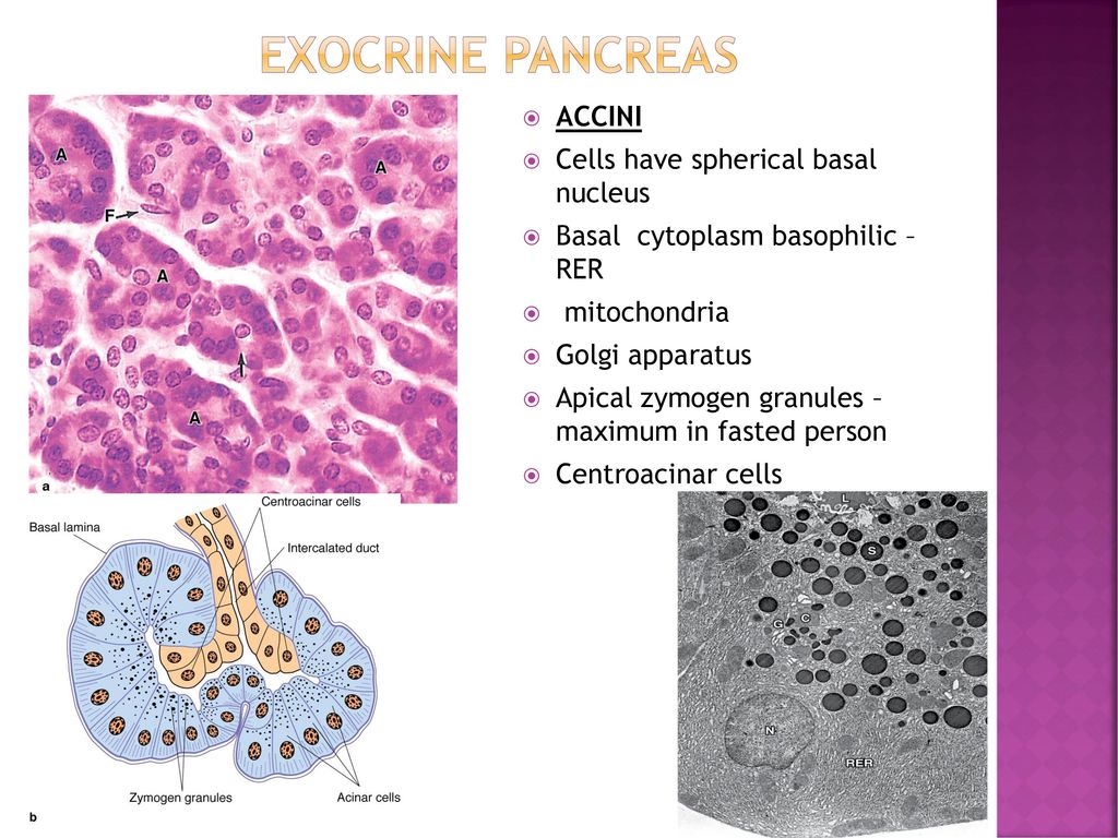


Histology Of Pancreas Ppt Download


2
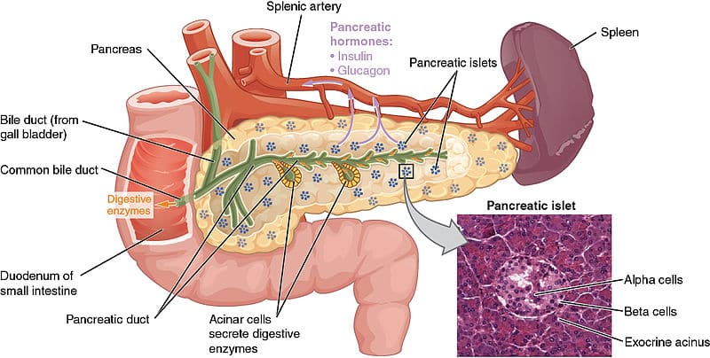
Pancreata from rodents and humans share many histological and functional similarities, but also display some potentially important differences ()The pancreas is invested by a variably thin connective tissue capsule and divided into lobules, which are formed from dense accumulations of exocrine glands that often surround the isletsIndividual lobules are defined byThe exocrine part of the pancreas has closely packed serous acini, similar to those of the digestive glands It secretes an enzyme rich alkaline fluid into the duodenum via the pancreatic ductThe pancreas is a large gland that has both exocrine and endocrine functions The majority of the pancreas consists of exocrine glands that produce about 15 liters of alkaline digestive enzymes daily, which is secreted directly into the duodenum The pancreas, also contains small endocrine cells found in clusters called islets of Langerhans
:watermark(/images/watermark_only.png,0,0,0):watermark(/images/logo_url.png,-10,-10,0):format(jpeg)/images/anatomy_term/pancreatic-acinar-cells/86g1HQPABGT8iQy0zPdrpg_Exocrine_cell_of_pancreas.png)


Pancreas Histology Exocrine Endocrine Parts Function Kenhub


Liver And Pancreas
The pancreasspecific proteome The pancreas is a composite organ with both exocrine and endocrine functions The exocrine compartment includes glandular cells that secrete enzymes to the gastrointestinal tract for digestion of food intake The endocrine function of the pancreas is based on the diffusely spread pancreatic endocrine cells, alsoIntralobular Duct (low columnar or cuboidal epithelium, nonEndocrine and exocrine pancreas develop at the same time Endocrine cells are first observed along the base of differentiating exocrine acini from weeks 1216 Compare and contrast microanatomy and blood supply of endocrine and exocrine portions of the pancreas


Department Of Pathology Ppt Video Online Download


Pin On Histology Slides
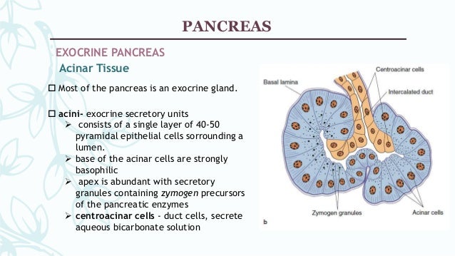
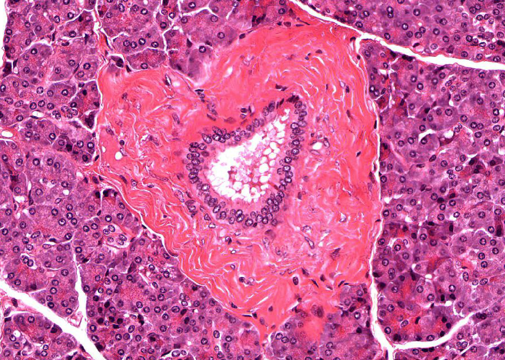
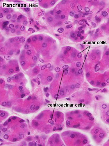
The pancreas is lumpy and glandular It comprises a head, neck, body, and tail, which points towards the spleen on the left side of the body The duodenum wraps around the head of the pancreas Exocrine Tissues The pancreatic tissue primarily comprises acini, which are clusters of secretory cells Visible as a circle of dark purplestaining Histology of the Pancreas Fig 31 This histologic section from the tail of the pancreas highlights the lobular architecture of the pancreatic parenchyma Several acini (exocrine) drain into small interlobular ducts ( short arrows ), which drain into a large duct ( long arrow ) The endocrine portion is characterized by several islets ofThis H&E section of the exocrine pancreas shows several of its characteristic features The exocrine cells show a strongly basophilic cytoplasm that represents the area occupied by the rough endoplasmic reticulum The apical side of the cells is filled with zymogen granules that contain a variety of digestive enzymes


The Pancreas Structure Role Pathology Study Com


Pancreatic Histology Exocrine Tissue
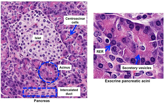
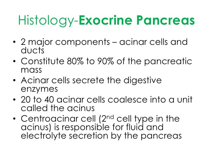
The pancreas is an organ containing two distinct populations of cells, the exocrine cells that secrete enzymes into the digestive tract, and the endocrine cells that secrete hormones into the bloodstream It arises from the endoderm as a dorsal and a ventral bud which fuse together to form the single organSecretory granules are located at the apex, adjacent to the lumen into which their contents will be released Centroacinar cells, located in the center of each acinus, form the beginning of the intralobular duct systemQualitative assessment of the number of islets of Langerhans and the degrees of exocrine pancreatic atrophy, fibrosis, and fat replacement was made for each pancreas Quantitative assessment of islet composition was performed in 15 of the 17 based on the immunochemical reactivity of each cell type Nondiabetic patients with cystic fibrosis in


Histology Gallbladder And Pancreas


Pancreas Histology Identifying Features With Labeled Slide Images Anatomylearner The Place To Learn Veterinary Anatomy Online
Exocrine pancreas, the portion of the pancreas that makes and secretes digestive enzymes into the duodenum This includes acinar and duct cells with associated connective tissue, vessels, and nerves The exocrine components comprise more than 95% of the pancreatic massVentral diverticulum gives the common bile duct, gallbladder, liver and ventral pancreatic anlage that becomes head of the pancreas;A brief review of the normal histology of the pancreas, as presented by the URMC Pathology IT Program


Pancreas Histology


Organisation Of The Human Pancreas In Health And In Diabetes Springerlink
HMB Pancreas Histology Liver Histology Gall Bladder Histology Renal System Histology exocrine pancreas consists of tubuloacinar glands single layer of pyramidal shaped cells forms the secretory acini (cells contain zymogen granules) Secretory duct pathway Intercalated Duct;The ducts that drain them secrete alkaline fluid Clusters of endocrine cells, called Islets of Langerhans, secrete hormones that regulate blood glucose metabolismHas islets of langerhans scattered throughout exocrine portion (secretory acini);


Pancreatic Phenotype A Histology Of The Exocrine Pancreatic Download Scientific Diagram


The Exocrine Pancreas Function Secretion Regulation
Unlike other exocrine glands, the intercalated ducts of the pancreas begin within the acini The cells that initiate the duct within the acini are called centroacinar cells because in section, the cells are visible as a lightly acidophilic squamous cell that occupies the sectioned lumen, ie, the center, of a pancreatic acinusThe pancreas is a long, slender organ, most of which is located posterior to the bottom half of the stomach (Figure 1791)Although it is primarily an exocrine gland, secreting a variety of digestive enzymes, the pancreas also has endocrine cells Its pancreatic islets—clusters of cells formerly known as the islets of Langerhans—secrete the hormones glucagon, insulin, somatostatin, and226 Pancreas Exocrine Pancreas View Virtual EM Slide In this low power electron micrograph, observe the organization of the acini, composed of acinar cells Within the acinar cells you will see the basal rough endoplasmic reticulum and the numerous secretory granules in the apical region of the cells, facing the small lumen of the acinus


Gastrointestinal Tract Pancreas Histology Embryology


2
The exocrine pancreas comprises the majority cell population and volume of the pancreas The exocrine pancreas is divided into lobules, which are separated by thin fibrovascular septa that coalesce with the pancreatic capsule which surrounds the organHistology At the histological level the pancreas is made up of compound glands in "bunch of grapes" fashion The pancreas has an exocrine and endocrine component The exocrine compnent is demonstrated above in 3D with acini in "cluster of grapes" formation subtended by a duct Courtesy Ashley Davidoff MD a06The pancreatic islets each contain four varieties of cells The alpha cell produces the hormone glucagon and makes up approximately percent of each islet, The pancreas 2, The mandate for this chapter is to review the anatomy and histology of the pancreas, Diabetes affects at least 30 million people world wide and despite the availability of
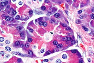


Histology Of The Pancreas Springerlink


Anatomy And Histology Of The Pancreas Pancreapedia
The exocrine pancreas has two types of cells The diagram illustrates the organization of the two major types of exocrine cells The acinar cells make powerful digestive enzymes (biological scissors) that can dissolve complex vegetables and meats, turning them into liquidsThe pancreas is formed from two basic tissue types termed the exocrine and endocrine pancreas Although the exocrine and endocrine pancreas share a similar anatomic location, for all intensive purposes they are histologically and physiologically distinct So too are the pathological processes which affect them Endocrine Histology Pancreas, Thymus and Pineal Body Full view of Pancreas Pancreatic islets of Langerhans are small nests of cells, arranged into curvilinear cords, scattered throughout the pancreas Islets are usually conspicuously paler (less intensely stained) than the acini (OR Exocrine Acinar Tissue) of the exocrine pancreas, but in any


Exocrine Gland Pancreas Histology Endocrine Gland Endocrine System Glands Texture Text Png Pngegg
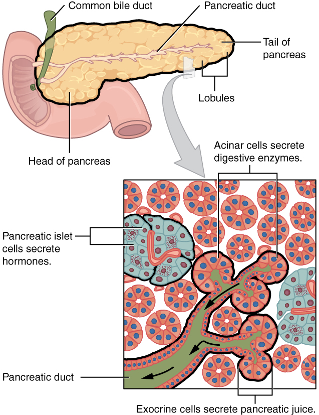


File 2424 Exocrine And Endocrine Pancreas Jpg Wikimedia Commons
Embryology of the pancreas develops from two outgrowths of the foregut;Pancreas is an exocrine organ The exocrine function involves producing digestive enzymes released into the small intestine We make more digestive enzymes than insulin so most of the cells are acinar cells that produce digestive enzymes The pancreatic isles, also called isles of Langerhan, produce insulin It is very difficult to


Scielo Brasil Comparative Evaluation Of Pancreatic Histopathology Of Rats Treated With Olanzapine Risperidone And Streptozocin Comparative Evaluation Of Pancreatic Histopathology Of Rats Treated With Olanzapine Risperidone And Streptozocin


Liver And Pancreas
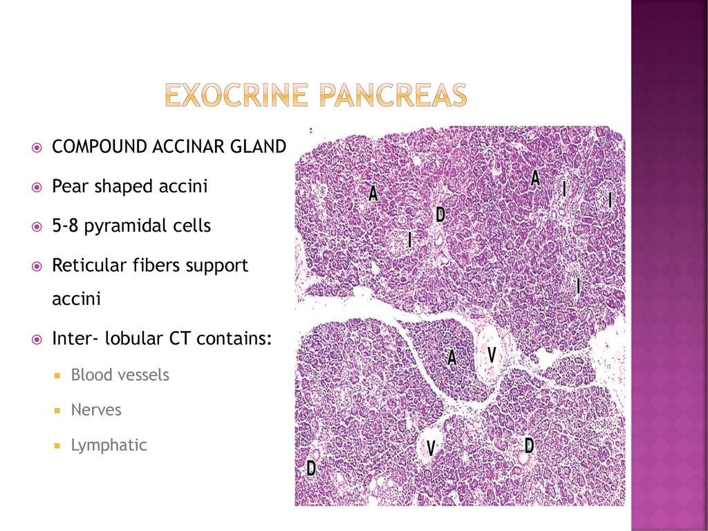


Histology Of Pancreas Ppt Download


01 07 15 Histology Of Tongue Liver Pancreas


Pancreas Sciencedirect


Histology Biol 4000 Digestive System Iii Lecture Notes 12c


Morphological Histological And Ultrastructural Studies On The Exocrine Pancreas Of Goose Sciencedirect


S 56 Histology Of Pancreas Exocrine Youtube
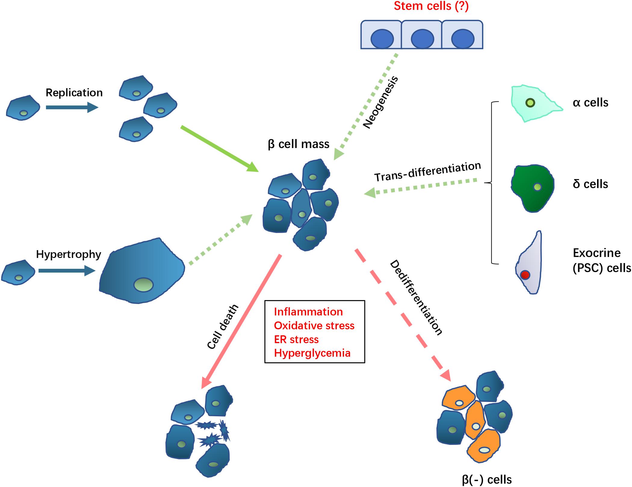


Frontiers Pathological Mechanisms In Diabetes Of The Exocrine Pancreas What S Known And What S To Know Physiology


Pancreatic Histology Hematoxylin Phloxin Saffron Stain 400 Acinar Download Scientific Diagram


Pancreas Histology


Pancreas Histology


Pancreas Histology Flashcards Quizlet


Pancreatic Histology Exocrine Tissue


Ha 235 Histology Accessory Digestive Glands


2


Pancreas Histology


Pancreatic Histology Exocrine Tissue


Pancreas Histology Boyar


Pancreatic Histology Endocrine Tissue


Anatomy And Histology Of The Pancreas Pancreapedia
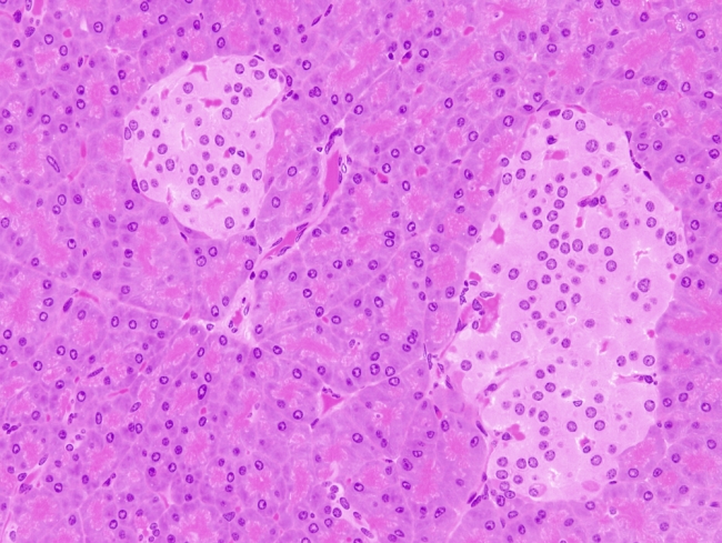


Animal Tissues Epithelium Pancreas Exocrine And Endocrine Gland Atlas Of Plant And Animal Histology


2


Pancreas


Normal Pancreas Pancreas Org


Basic Histology Exocrine Pancreas


Endocrine Pancreas


Pituitary Gland


Pancreas Objectives The Student Should Be Able To Describe 1 The Endocrine Part Of The Pancreas Within The Exocrine Part 2 The Histological Features Ppt Download


Exocrine Pancreas An Overview Sciencedirect Topics


Anatomy And Histology Of The Pancreas Pancreapedia


Pancreas Slide Wsu 2 060


Pathology Of The Exocrine Pancreas Tyler Verdun Pgy
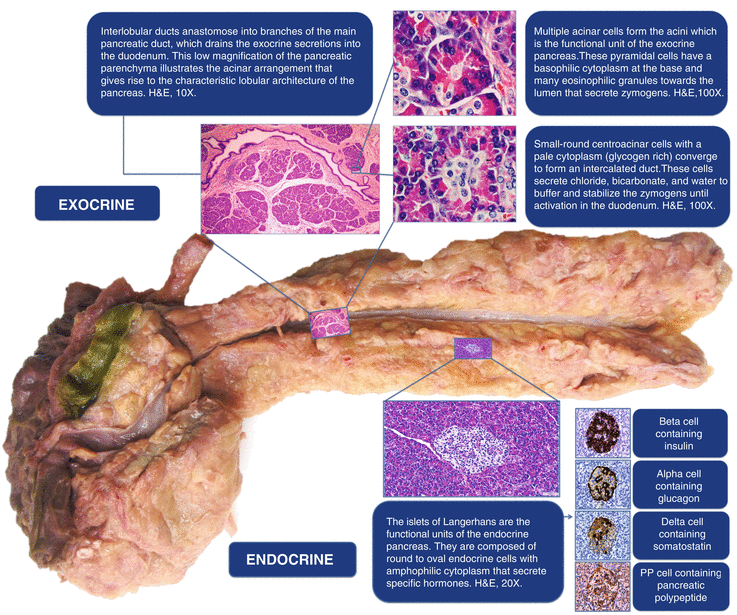


Histology Of The Pancreas Springerlink


The Roles Of Calcium And Atp In The Physiology And Pathology Of The Exocrine Pancreas Physiological Reviews


Pancreas Histology Osmosis


Exocrine Pancreas Flashcards Quizlet


Histology Of Pancreas Youtube
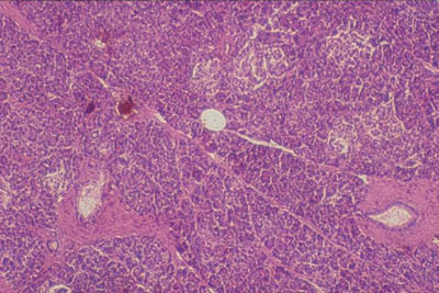


Histology World Histology Fact Sheet Pancreas
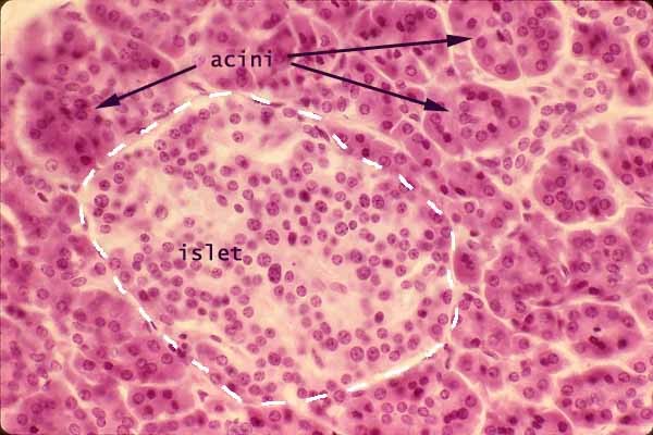


Histology At Siu


Digestive The Histology Guide


Endocrine And Exocrine Pancreas In 21 Pancreas Histology Slides Endocrine System


Histoquarterly Pancreas Histology Blog Medical School Stuff Histology Slides Endocrine System


Chapter 16 Page 3 Histologyolm


Light Micrograph Of Sections Of Exocrine Pancreas A Control Group B C Download Scientific Diagram
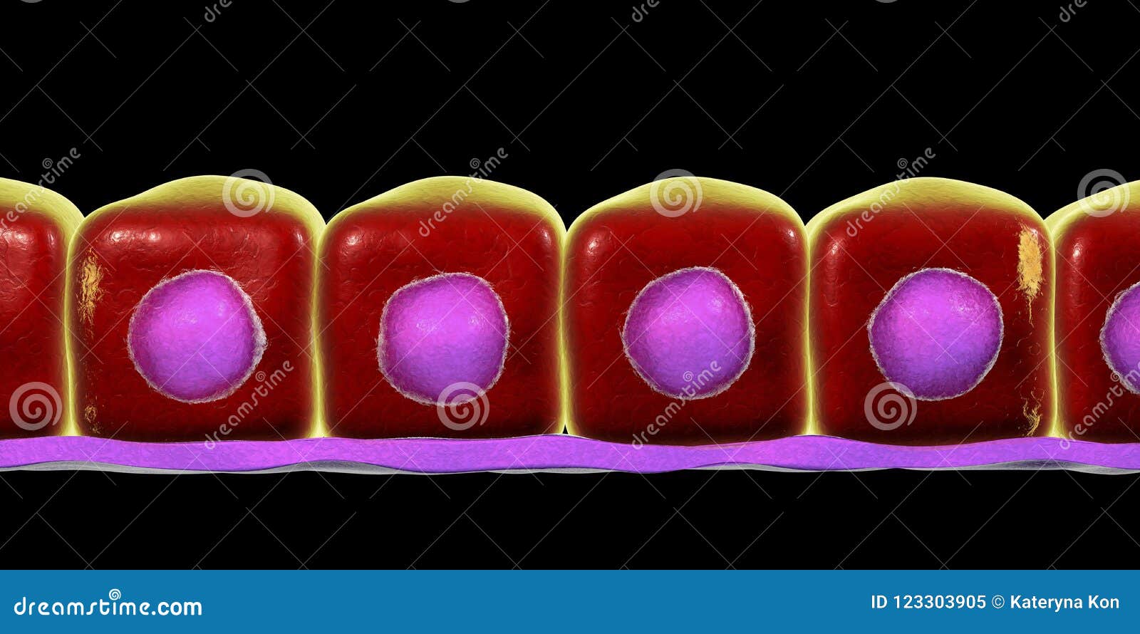


Simple Cuboidal Epithelium Stock Illustration Illustration Of Exocrine


Pancreas Sciencedirect


Chapter 15 Page 7 Histologyolm 4 0


01 07 15 Histology Of Tongue Liver Pancreas


Pancreas Histology Osmosis
:background_color(FFFFFF):format(jpeg)/images/article/en/pancreas-histology/VMvxP28VGT55N9WvU3WgQ_mbuhIx2qC3HVaYNH69w_Body_of_Pancreas.png)


Pancreas Histology Exocrine Endocrine Parts Function Kenhub


Acinar


Histology Of The Pancreas Endocrine And Exocrine Youtube
:watermark(/images/watermark_5000_10percent.png,0,0,0):watermark(/images/logo_url.png,-10,-10,0):format(jpeg)/images/overview_image/1881/CPsIC67xuz6pLt6JhAM6Wg_pancreas-histology_english.jpg)


Pancreas Histology Exocrine Endocrine Parts Function Kenhub


Pancreatic Islets Wikipedia


Histoquarterly Pancreas Histology Blog


Dictionary Normal Pancreas The Human Protein Atlas


Anatomy And Histology Of The Pancreas Pancreapedia


Histology Of Mouse Pancreas Stained By Hematoxylin Eosin Normal Download Scientific Diagram


Pancreas Histology Draw It To Know It
:watermark(/images/watermark_5000_10percent.png,0,0,0):watermark(/images/logo_url.png,-10,-10,0):format(jpeg)/images/overview_image/1059/SXYoNKo9I26Aroj0YPbuHg_pancreatic-duct-system_english.jpg)


Pancreas Histology Exocrine Endocrine Parts Function Kenhub


Pancreatic Histology Endocrine Tissue


1 Schematic Histology Of The Pancreas Pancreatic Islets Are Download Scientific Diagram


Ha 235 Histology Accessory Digestive Glands


Pancreas Histology Histological Section Through A Wild Type Mouse Download Scientific Diagram


Pancreatic Histology Exocrine Tissue


9 Pancreas Ideas Pancreas Anatomy And Physiology Histology Slides


Pancreas Histology
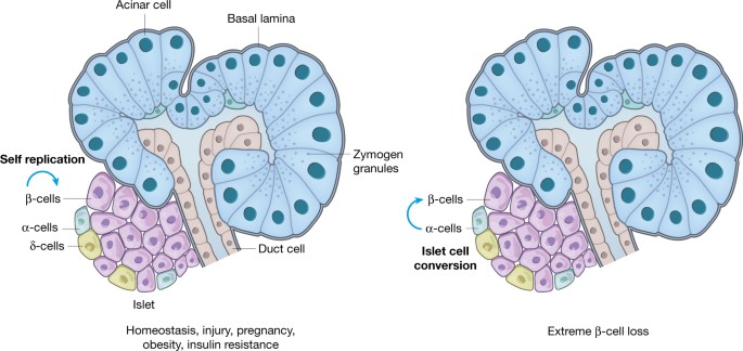


Pancreas Regeneration Nature


Human Structure Virtual Microscopy


File Pancreas Histology 002 Jpg Embryology


Solved Pancreas Histology Practice Name This Group Of Cells Chegg Com


Pathology Of The Pancreas Basicmedical Key


File Pancreas Histology 102 Jpg Embryology


Glandular Tissue The Histology Guide


Exocrine Gland Wikipedia


Liver And Pancreas


Informacion Adicional Del Pancreas
コメント
コメントを投稿